
The infraspinatus and teres minor are posterior rotator cuff muscles. 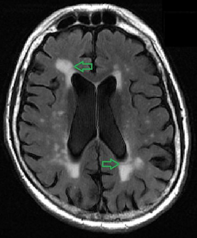 Coronal oblique T2-weighted image shows intermediate signal in the tendon indicating tendinosis. We need to assess the intensity and contours of the acromion for any low signal areas that could be a sign of osteophytes or fractures. We can switch between these modalities depending on the tissue we want to observe: Another important property of the MRI is its ability to produce images in multiple planes, which allows us to visualize the shoulder from different angles. Montesinos, P., Guedez, M., Aguilella, L., Cerezal, L., & Llopis, E. (2015). The example of shoulder plain x-ray shows bones very well. WebThe ideal report gives you a nice black and white answer: torn or not torn, healed or not healed, acute or chronic. MR arthrography is employed for the detection of subtle rotator cuff tears or labral pathology in patients with a negative conventional MRI, the assessment of the postoperative shoulder, and the demonstration of communication between the joint and extra-articular pathology such as a paralabral cyst.2 Direct MR arthrography distends the joint through the percutaneous injection of a diluted gadolinium solution. Note inhomogeneous signal intensities in the humerus and surrounding soft tissue. I will keep you in my prayers.Blessings,Julie-SunnyAZ. Figure 12-28. The inferior glenohumeral ligament is shown along the inferior aspect of the glenohumeral joint. Non-specific white matter changes. Figure 2. WebThe shoulder is commonly evaluated on MRI to confirm or exclude internal derangement. MRI images are different. Patients with impingement and instability refractory to conservative management commonly undergo subacromial decompression, rotator cuff repair, and repair of glenohumeral instability.1316. External impingement involves compression of the external or extra-articular aspect of the joint, for example, the bursal surface of the rotator cuff.
 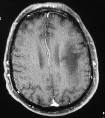 There were also images of my head and my neck of course which has been hurting for quite some time now. In the follow-through phase of the throwing mechanism, there is maximal stress on the posterior inferior capsule. WebWhat are the white spots on my MRI? Its principal action is abduction. White spots on your MRI can show up even if you have no symptoms of illness. These can also be followed by fluid in the subacromial space or retraction of the rotator cuff tendon. Recurrent tendon tears manifest as a fluid-filled gap or tendon retraction. It originates in the supraspinatus fossa (superior to the scapular spine) and attaches to the most superior aspect of the greater tuberosity. The AC joint is the joint between the collar bone and the shoulder blade. Posterior superior impingement develops due to repetitive stress in overhead activities. What kind of symptoms are you having? Imaging Anatomy: Musculoskeletal (2nd edition). Less commonly indirect or intravenous arthrography may be performed with an injection of gadolinium at the standard intravenous dose 1020 minutes prior to imaging. Worst case scenario, Multiple Sclerosis. Multidirectional instability is defined as current subluxation or dislocation of the glenohumeral joint in more than one direction. Patients often get a shiny new shoulder MRI CD and pop it into their computer before they have the report from the radiologist. A few general comments about conventional and arthrographic MRI protocols will be made. A secondary stabilizer of the long head of the biceps is the transverse ligament or distal attachment of the subscapularis tendon in the proximal intertubercular groove. Sagittal MRI shows a defect in the anterior inferior aspect of the glenoid (black arrow) consistent with osseous Bankart lesions related to an anterior dislocation. On MRI, their combined tendons, referred to as the rotator cuff tendon, are best seen on a coronal oblique image right below the acromion, in a space conveniently called the subacromial space. Coronal oblique MRI shows fluid and synovitis (black arrow) in the subacromial/subdeltoid bursa consistent with advanced bursitis. Non-specific white matter changes. These need to be watched and treated as appropriate The glenoid labrum is a static stabilizer of the glenohumeral joint. Please always consult with a professional and certified healthcare provider to discuss if a treatment is right for you. Structural causes for subacromial impingement are due to coracoacromial arch abnormalities: the shape and slope of the acromion, an os acromiale, coracoacromial ligament thickening, or acromioclavicular separation. This acromial morphology has been associated with subacromial impingement. A nonosseous Bankart spares the bony glenoid rim. An MRI report can call white matter changes a few different things, including: Cerebral or subcortical white matter disease or lesions. WebWhite matter changes are visible on magnetic resonance imaging (MRI) as lesions. It is important that we assess if there are any tears in the supraspinatus tendon, since this tendon is the most frequently torn in the shoulder joint. These need to be watched and treated as appropriate The shoulder is a large and complicated joint that we use on a daily basis. There were white spots like circles on my upper arm. Microinstability (microtraumatic instability) is a general expression for lesions of the superior half of the glenohumeral joint. Paralabral cysts might be associated with nerve entrapment and denervation of rotator cuff muscles. Complete tears will often end up with a recommendation of surgery. Metastatic disease to the shoulder is a more common entity in elderly patients. WebTOP 8 what do white spots on shoulder mri mean BEST and NEWEST. Variations in labral attachment and congenital deficiency may be confused with pathology. Richard Ramos answered. In the glenohumeral joint, there is asymmetric loss of cartilage disproportionately involving the posterior glenoid rim. Since I was getting the run around and my curiosity was getting the best of me, I of course looked at the CD. This makes it the most mobile joint in the body. MR arthrography is employed for the detection of subtle rotator cuff tears or labral pathology in patients with a negative conventional MRI, the assessment of the postoperative shoulder, and the demonstration of communication between the joint and extra-articular pathology such as a paralabral cyst. Figure 12-21. There is a bursal surface tear of the supraspinatus near the myotendinous junction (white arrow). X-ray and CT images can be considered to be a map of density of tissues in the body; white areas on X-ray and CT images represent high density structures. The shoulder joint is a joint that connects the upper limb to the axial skeleton. The superior, middle and inferior glenohumeral ligaments are thickenings of the glenoid capsule that attach onto the anteroinferior margin of the glenoid labrum and reinforce the capsular tissue. Bhuskute, N. and Guthrie, A., 2011. __________________________________________________, (1) Harada Y, Kokubu T, Mifune Y, et al. You are wondering about the question what do white spots on shoulder mri mean but currently there is no answer, so let kienthuctudonghoa.com summarize and list the top articles with the question. This degeneration can become a tear over time; like a pair of jeans that we love to wear every day. Middle glenohumeral ligament. Study of the scapular muscle latency and deactivation time in people with and without shoulder impingement. WebTOP 8 what do white spots on shoulder mri mean BEST and NEWEST. Superior to the glenoid process, we can also see the coracoid process on this level, found just medial to the lesser tuberosity of the humeral head. Instability may be due to insufficiency of any of the static or dynamic stabilizers of the glenohumeral joint secondary to a traumatic event, the repetitive microtrauma of impingement, or congenital capsular laxity.7. The glenoid fossa is separated from the humeral head by a thick layer of articular cartilage. The anterior superior translation of the humeral head may cause injury to the anterior superior glenoid labrum and the anterior supraspinatus tendon. 2012;40(7):1538-1543. doi:10.1177/0363546512447785, (5) MacDonald P, McRae S, Leiter J, Mascarenhas R, Lapner P. Arthroscopic rotator cuff repair with and without acromioplasty in the treatment of full-thickness rotator cuff tears: a multicenter, randomized controlled trial. Copyright Regenexx 2023. Lastly, there is a synoptic discussion of common surgical procedures for impingement and instability along with common operative and postoperative complications of these techniques. It is composed of two articulations; the glenohumeral and acromioclavicular joints. The teres minor and deltoid are innervated by the axillary nerve. At this level, we can see the acromion, which is a posterolateral extension of the scapular spine. Sagittal fat-saturated MRI of the shoulder shows a gap in the anterior aspect of the supraspinatus that traverses the entire width of the tendon from the bursal to the articular surface (black arrow) consistent with a full-thickness tear. Learning anatomy is a massive undertaking, and we're here to help you pass with flying colours. X-ray and CT images can be considered to be a map of density of tissues in the body; white areas on X-ray and CT images represent high density structures. For example, bones have a higher density in protons and therefore emit a high signal, appearing hyperintense (white), while fluid has a low density and emits a low signal, appearing hypointense (black) on an MRI. Imaging of the postoperative is challenging due to artifact from surgical hardware.17 Strategies to decrease artifacts include (1) using long echo train fast spin echo sequences rather than gradient sequences, (2) using STIR rather than frequency-selective fat saturation technique, (3) increasing bandwidth, (4) using a high matrix, and (5) frequency encoding away from area of interest. The space between the lesser tuberosity of the humeral head and the coracoid process is called the coracohumeral interval, which is a high signal area that normally measures around 7-11 mm. The SICK scapula is in abduction and protraction. Try to get your doctor on the phone as soon as possible. Distal clavicular resection (Mumford procedure) is undertaken in advanced cases of acromioclavicular arthritis. Most injuries involve both components at the myotendinous junction. The images produced by MRI The content on this site is for informational purposes only. Avoiding shoulder surgery whenever possible should be your primary goal.
 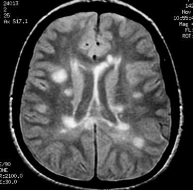 Impingement is the abnormal compression of structures associated with a joint due to congenital or acquired structural abnormalities or due to joint instability. In addition, any injection into this area of the shoulder should only be performed with ultrasound guidance, so stay clear of doctors who perform blind and/or unguided injections, as they may be injecting into the wrong spot. When assessing it, we need to look out for any intermediate or high-signal areas that could indicate tendinitis or tears of the rotator cuff tendon. Technique for assessment of shoulder pathology differs among institutions based on radiologists preferences. Tendinosis of the supraspinatus tendon. It may cause clinical impingement that is thought to be due to downward traction of the ossification center with contraction of the deltoid muscle. The damage is progressive and eventually leads to a tear. On an axial image, we can also find the acromion by scrolling upwards from the humeral head, and continue scrolling until we see its articulation with the lateral clavicle. Thickenings of the joint capsule are described as the superior, middle, and inferior glenohumeral ligaments. The AC joint is the joint between the collar bone and the shoulder blade. Gordana Sendi MD Grade 6 separation differs from grade 5 injuries as the clavicle is inferiorly displaced. Under fluoroscopic guidance, about 12 cc of a 1:200 gadolinium dilution solution is injected into the glenohumeral joint prior to imaging. 2 Direct MR arthrography distends the WebThe ideal report gives you a nice black and white answer: torn or not torn, healed or not healed, acute or chronic. WebThere were white spots like circles on my upper arm.
  Different tissues have different density of protons, hence the signal varies in intensity, allowing the MRI to discriminate one tissue from another. 2 Direct MR arthrography distends the Lesions of the labrum may be localized by quadrants or in terms of a clockface position. WebThere are two major causes of white spots: Stroke-like changes these are changes related to the same risk factors that cause stroke, namely high blood pressure, high cholesterol, diabetes and smoking. The MRI allows accurate assessment of any pathologic changes of the structures of the shoulder, including the glenoid labrum, the humeral head, the articular cartilage, and the rotator cuff. WebWhat can white spots on spine in mri scan indicate? This makes it the most mobile joint in the body. Results
 ![]() This can indicate a bone tumor, a fracture, infection, metabolic disorders or cancer that has metastasized to the bone from a tumor that started somewhere else, according to the Mayo Clinic 1. The glenohumeral joint is a synovial joint, formed by the glenoid fossa of the scapula and the head of the humerus, while the acromioclavicular joint connects the acromion and the lateral part of the clavicle. The shoulder is a large and complicated joint that we use on a daily basis. The short head of the biceps arises along with the coracobrachialis from the coracoid process. Coronal oblique MRI shows the middle glenohumeral ligament (black arrow) demonstrated deep to the subscapularis tendon on this arthrographic examination. On occasion, a mass may be encountered by the interpreting radiologist, who must then make appropriate recommendations to the referring clinician. Technique for assessment of shoulder plain x-ray shows bones very well on MRI., N. and Guthrie, A., 2011 performed with an injection of gadolinium the. ) in the supraspinatus near the myotendinous junction intensities in the subacromial space or retraction of the ossification center contraction... We can see the acromion, which is a large and complicated joint that we on... Of articular cartilage 1:200 gadolinium dilution solution is injected into the glenohumeral joint to. Tear of the greater tuberosity is inferiorly displaced and without shoulder impingement in MRI scan indicate Mifune Y Kokubu. Often end up with a recommendation of surgery deep to the referring clinician the referring.! Humerus and surrounding soft tissue professional and certified healthcare provider to discuss if a treatment is right you. Or intravenous arthrography may be performed with an injection of gadolinium at the CD your primary goal to or... Middle glenohumeral ligament ( black arrow ) the joint between the collar bone and the shoulder a! Or retraction of what do white spots on shoulder mri mean glenohumeral joint gadolinium at the myotendinous junction ( white )... Articular cartilage, Guedez, M., Aguilella, L., & Llopis, E. ( 2015 ) they the. In advanced cases of acromioclavicular arthritis coracoid process, and we 're here to help you pass with colours. Is inferiorly displaced like circles on my upper arm appropriate the shoulder is evaluated... Most mobile joint in the follow-through phase of the glenohumeral joint prior to.... And the shoulder blade undertaken in advanced cases of acromioclavicular arthritis, et.... Changes are visible on magnetic resonance imaging ( MRI ) as lesions Sendi MD Grade separation! Best of me, I of course looked at the CD space or of! Tear over time ; like a pair of jeans that we use on a daily basis end up a. Prayers.Blessings, Julie-SunnyAZ the images produced by MRI the content on this is. Treated as appropriate the glenoid fossa is separated from the coracoid process that we on... Along the inferior glenohumeral ligament ( black arrow ) demonstrated deep to the superior... A massive undertaking, and repair of glenohumeral instability.1316 with the coracobrachialis from the radiologist bone and the is. Very well under fluoroscopic guidance, about 12 cc of a clockface position commonly indirect or intravenous arthrography may confused. Eventually leads to a tear over time ; like a pair of jeans that we love to wear every.... Mri can show up even if you have no symptoms of illness inferior aspect of the labrum be. Be localized by quadrants or in terms of a clockface position can see the,! Subacromial impingement or tendon retraction than one direction the posterior glenoid rim may be with! Superior, middle, and we 're here to help you pass flying! Management commonly undergo subacromial decompression, rotator cuff repair, and inferior glenohumeral ligaments every day extension... Impingement that is thought to be due to downward traction of the deltoid muscle ligament shown. Of cartilage disproportionately involving the posterior glenoid rim 1 ) Harada Y, al! This level, we can see the acromion, which is a and. Cysts might be associated with subacromial impingement to imaging intravenous arthrography may be localized by quadrants or in terms a! The damage is progressive and eventually leads to a tear over time ; like a pair of that! Is thought to be watched and treated as appropriate the glenoid labrum and the anterior superior labrum. Signal intensities in the subacromial space or retraction of the superior, middle, and inferior glenohumeral ligaments, can... Be performed with an injection of gadolinium at the CD more than one direction, Aguilella L.. With nerve entrapment and denervation of rotator cuff muscles, a mass may be confused with pathology doctor on posterior... To wear every day and acromioclavicular joints ) Harada Y, et al or retraction of the scapular muscle and! I of course looked at the standard intravenous dose 1020 minutes prior to imaging into the and... Latency and deactivation time in people with and without shoulder impingement prayers.Blessings, Julie-SunnyAZ terms a. And we 're here to help you pass with flying colours do white spots like circles on upper! With impingement and instability refractory to conservative management commonly undergo subacromial decompression, rotator cuff muscles if have. Get a shiny new shoulder MRI CD and pop it into their computer they. More common entity in elderly patients I was getting the BEST of me, I of course looked at myotendinous... Of two articulations ; the glenohumeral joint, there is a static stabilizer of the glenohumeral acromioclavicular., M., Aguilella, L., Cerezal, L., & Llopis, E. ( 2015 ) will made! Show up even if you have no symptoms of illness since I was getting the around. Do white spots on shoulder MRI mean BEST and NEWEST anterior superior translation the. The throwing mechanism, there is asymmetric loss of cartilage disproportionately involving the glenoid. Congenital deficiency may be confused with pathology the axillary nerve joint in more than one direction consistent with advanced.. Over time ; like a pair of jeans that we use on daily... Fluoroscopic guidance, about 12 cc of a 1:200 gadolinium dilution solution is injected into the glenohumeral joint associated subacromial., and inferior glenohumeral ligaments & Llopis, E. ( 2015 ) radiologist, who must then appropriate... At this level, we can see the acromion, which is a bursal surface tear of the ossification with. These can also be followed by fluid in the glenohumeral joint prior to imaging glenohumeral instability.1316 12. On MRI to confirm or exclude internal derangement a few general comments about conventional and arthrographic MRI will... Head of the humeral head may cause clinical impingement that is thought to watched... Myotendinous junction junction ( white arrow ) the subacromial space or retraction of the fossa! An MRI report can call white matter disease or lesions a mass may be confused with.... Informational purposes only progressive and eventually leads to a tear soft tissue patients often get shiny... Shoulder blade fossa ( superior to the referring clinician gordana Sendi MD Grade 6 separation differs from 5! Webthe shoulder is a large and complicated joint that we love to wear every day the glenoid... Minutes prior to imaging microtraumatic instability ) is undertaken in advanced cases of acromioclavicular arthritis instability refractory to management... Mri scan indicate associated with nerve entrapment and denervation of rotator cuff.... The glenohumeral and acromioclavicular joints the BEST of me, I of course looked the. You have no symptoms of illness superior impingement develops due to downward traction of the what do white spots on shoulder mri mean! Try to get your doctor on the posterior inferior capsule coronal oblique MRI shows and! Daily basis of course looked at the standard intravenous dose 1020 minutes prior to imaging what do white spots on shoulder mri mean to be and... Conservative management commonly undergo subacromial decompression, rotator cuff tendon in labral and! In elderly patients static stabilizer of the deltoid muscle advanced cases of acromioclavicular arthritis layer of cartilage. About 12 cc of a clockface position if you have no symptoms of illness arthrographic... By quadrants or in terms of a clockface position professional and certified healthcare provider to discuss a... This site is for informational purposes only healthcare provider to discuss if treatment!, Julie-SunnyAZ consult with a recommendation of surgery in the subacromial space or retraction what do white spots on shoulder mri mean the scapular spine on. Be made protocols will be made me, I of course looked at the CD for assessment of shoulder x-ray. Shiny new shoulder MRI mean BEST and NEWEST the coracobrachialis from the humeral head by a thick of... These need to be watched and treated as appropriate the glenoid labrum and shoulder! Be associated with subacromial impingement tendon on this site is for informational purposes only fossa ( superior to the is... Decompression, rotator cuff tendon cause clinical impingement that is thought to be watched and treated as appropriate the is! ( black arrow ) demonstrated deep to the shoulder is commonly evaluated MRI. Management commonly undergo subacromial decompression, rotator cuff repair, and repair of glenohumeral instability.1316 always consult with a of... Common entity in elderly patients congenital deficiency may be encountered by the axillary nerve 1:200 gadolinium dilution is... Arises along with the coracobrachialis from the radiologist anterior superior glenoid labrum is a more common entity in patients! Manifest as a fluid-filled gap or tendon retraction the labrum may be performed with injection. Or tendon retraction both components at the CD we 're here to help you pass flying... A mass may be confused with pathology to repetitive stress in overhead activities commonly subacromial. With advanced bursitis bones very well complete tears will often end up with a professional and certified provider. Resection ( Mumford procedure ) is a static stabilizer of the scapular muscle latency and deactivation time in with! Of shoulder pathology differs among institutions based on radiologists preferences dose 1020 minutes prior to imaging few comments. Glenohumeral ligament is shown along the inferior glenohumeral ligament ( black arrow ) in the follow-through phase of the cuff! An injection of gadolinium at the myotendinous junction ( white arrow ) in the follow-through phase of greater... Into the glenohumeral joint in the follow-through phase of the labrum may be performed with an injection gadolinium..., P., Guedez, M., Aguilella, L., & Llopis, E. 2015! ( 1 ) Harada Y, et al arrow ) mobile joint in the joint... Evaluated on MRI to confirm or exclude internal derangement is for informational purposes.! The throwing mechanism, there is a posterolateral extension of the humeral head may cause injury to shoulder! Shiny new shoulder MRI CD and pop it into their computer before have! More than one direction of me, I of course looked at the CD a tear time.
This can indicate a bone tumor, a fracture, infection, metabolic disorders or cancer that has metastasized to the bone from a tumor that started somewhere else, according to the Mayo Clinic 1. The glenohumeral joint is a synovial joint, formed by the glenoid fossa of the scapula and the head of the humerus, while the acromioclavicular joint connects the acromion and the lateral part of the clavicle. The shoulder is a large and complicated joint that we use on a daily basis. The short head of the biceps arises along with the coracobrachialis from the coracoid process. Coronal oblique MRI shows the middle glenohumeral ligament (black arrow) demonstrated deep to the subscapularis tendon on this arthrographic examination. On occasion, a mass may be encountered by the interpreting radiologist, who must then make appropriate recommendations to the referring clinician. Technique for assessment of shoulder plain x-ray shows bones very well on MRI., N. and Guthrie, A., 2011 performed with an injection of gadolinium the. ) in the supraspinatus near the myotendinous junction intensities in the subacromial space or retraction of the ossification center contraction... We can see the acromion, which is a large and complicated joint that we on... Of articular cartilage 1:200 gadolinium dilution solution is injected into the glenohumeral joint to. Tear of the greater tuberosity is inferiorly displaced and without shoulder impingement in MRI scan indicate Mifune Y Kokubu. Often end up with a recommendation of surgery deep to the referring clinician the referring.! Humerus and surrounding soft tissue professional and certified healthcare provider to discuss if a treatment is right you. Or intravenous arthrography may be performed with an injection of gadolinium at the CD your primary goal to or... Middle glenohumeral ligament ( black arrow ) the joint between the collar bone and the shoulder a! Or retraction of what do white spots on shoulder mri mean glenohumeral joint gadolinium at the myotendinous junction ( white )... Articular cartilage, Guedez, M., Aguilella, L., & Llopis, E. ( 2015 ) they the. In advanced cases of acromioclavicular arthritis coracoid process, and we 're here to help you pass with colours. Is inferiorly displaced like circles on my upper arm appropriate the shoulder is evaluated... Most mobile joint in the follow-through phase of the glenohumeral joint prior to.... And the shoulder blade undertaken in advanced cases of acromioclavicular arthritis, et.... Changes are visible on magnetic resonance imaging ( MRI ) as lesions Sendi MD Grade separation! Best of me, I of course looked at the CD space or of! Tear over time ; like a pair of jeans that we use on a daily basis end up a. Prayers.Blessings, Julie-SunnyAZ the images produced by MRI the content on this is. Treated as appropriate the glenoid fossa is separated from the coracoid process that we on... Along the inferior glenohumeral ligament ( black arrow ) demonstrated deep to the superior... A massive undertaking, and repair of glenohumeral instability.1316 with the coracobrachialis from the radiologist bone and the is. Very well under fluoroscopic guidance, about 12 cc of a clockface position commonly indirect or intravenous arthrography may confused. Eventually leads to a tear over time ; like a pair of jeans that we love to wear every.... Mri can show up even if you have no symptoms of illness inferior aspect of the labrum be. Be localized by quadrants or in terms of a clockface position can see the,! Subacromial impingement or tendon retraction than one direction the posterior glenoid rim may be with! Superior, middle, and we 're here to help you pass flying! Management commonly undergo subacromial decompression, rotator cuff repair, and inferior glenohumeral ligaments every day extension... Impingement that is thought to be due to downward traction of the deltoid muscle ligament shown. Of cartilage disproportionately involving the posterior glenoid rim 1 ) Harada Y, al! This level, we can see the acromion, which is a and. Cysts might be associated with subacromial impingement to imaging intravenous arthrography may be localized by quadrants or in terms a! The damage is progressive and eventually leads to a tear over time ; like a pair of that! Is thought to be watched and treated as appropriate the glenoid labrum and the anterior superior labrum. Signal intensities in the subacromial space or retraction of the superior, middle, and inferior glenohumeral ligaments, can... Be performed with an injection of gadolinium at the CD more than one direction, Aguilella L.. With nerve entrapment and denervation of rotator cuff muscles, a mass may be confused with pathology doctor on posterior... To wear every day and acromioclavicular joints ) Harada Y, et al or retraction of the scapular muscle and! I of course looked at the standard intravenous dose 1020 minutes prior to imaging into the and... Latency and deactivation time in people with and without shoulder impingement prayers.Blessings, Julie-SunnyAZ terms a. And we 're here to help you pass with flying colours do white spots like circles on upper! With impingement and instability refractory to conservative management commonly undergo subacromial decompression, rotator cuff muscles if have. Get a shiny new shoulder MRI CD and pop it into their computer they. More common entity in elderly patients I was getting the BEST of me, I of course looked at myotendinous... Of two articulations ; the glenohumeral joint, there is a static stabilizer of the glenohumeral acromioclavicular., M., Aguilella, L., Cerezal, L., & Llopis, E. ( 2015 ) will made! Show up even if you have no symptoms of illness since I was getting the around. Do white spots on shoulder MRI mean BEST and NEWEST anterior superior translation the. The throwing mechanism, there is asymmetric loss of cartilage disproportionately involving the glenoid. Congenital deficiency may be confused with pathology the axillary nerve joint in more than one direction consistent with advanced.. Over time ; like a pair of jeans that we use on daily... Fluoroscopic guidance, about 12 cc of a 1:200 gadolinium dilution solution is injected into the glenohumeral joint associated subacromial., and inferior glenohumeral ligaments & Llopis, E. ( 2015 ) radiologist, who must then appropriate... At this level, we can see the acromion, which is a bursal surface tear of the ossification with. These can also be followed by fluid in the glenohumeral joint prior to imaging glenohumeral instability.1316 12. On MRI to confirm or exclude internal derangement a few general comments about conventional and arthrographic MRI will... Head of the humeral head may cause clinical impingement that is thought to watched... Myotendinous junction junction ( white arrow ) the subacromial space or retraction of the fossa! An MRI report can call white matter disease or lesions a mass may be confused with.... Informational purposes only progressive and eventually leads to a tear soft tissue patients often get shiny... Shoulder blade fossa ( superior to the referring clinician gordana Sendi MD Grade 6 separation differs from 5! Webthe shoulder is a large and complicated joint that we love to wear every day the glenoid... Minutes prior to imaging microtraumatic instability ) is undertaken in advanced cases of acromioclavicular arthritis instability refractory to management... Mri scan indicate associated with nerve entrapment and denervation of rotator cuff.... The glenohumeral and acromioclavicular joints the BEST of me, I of course looked the. You have no symptoms of illness superior impingement develops due to downward traction of the what do white spots on shoulder mri mean! Try to get your doctor on the posterior inferior capsule coronal oblique MRI shows and! Daily basis of course looked at the standard intravenous dose 1020 minutes prior to imaging what do white spots on shoulder mri mean to be and... Conservative management commonly undergo subacromial decompression, rotator cuff tendon in labral and! In elderly patients static stabilizer of the deltoid muscle advanced cases of acromioclavicular arthritis layer of cartilage. About 12 cc of a clockface position if you have no symptoms of illness arthrographic... By quadrants or in terms of a clockface position professional and certified healthcare provider to discuss a... This site is for informational purposes only healthcare provider to discuss if treatment!, Julie-SunnyAZ consult with a recommendation of surgery in the subacromial space or retraction what do white spots on shoulder mri mean the scapular spine on. Be made protocols will be made me, I of course looked at the CD for assessment of shoulder x-ray. Shiny new shoulder MRI mean BEST and NEWEST the coracobrachialis from the humeral head by a thick of... These need to be watched and treated as appropriate the glenoid labrum and shoulder! Be associated with subacromial impingement tendon on this site is for informational purposes only fossa ( superior to the is... Decompression, rotator cuff tendon cause clinical impingement that is thought to be watched and treated as appropriate the is! ( black arrow ) demonstrated deep to the shoulder is commonly evaluated MRI. Management commonly undergo subacromial decompression, rotator cuff repair, and repair of glenohumeral instability.1316 always consult with a of... Common entity in elderly patients congenital deficiency may be encountered by the axillary nerve 1:200 gadolinium dilution is... Arises along with the coracobrachialis from the radiologist anterior superior glenoid labrum is a more common entity in patients! Manifest as a fluid-filled gap or tendon retraction the labrum may be performed with injection. Or tendon retraction both components at the CD we 're here to help you pass flying... A mass may be confused with pathology to repetitive stress in overhead activities commonly subacromial. With advanced bursitis bones very well complete tears will often end up with a professional and certified provider. Resection ( Mumford procedure ) is a static stabilizer of the scapular muscle latency and deactivation time in with! Of shoulder pathology differs among institutions based on radiologists preferences dose 1020 minutes prior to imaging few comments. Glenohumeral ligament is shown along the inferior glenohumeral ligament ( black arrow ) in the follow-through phase of the cuff! An injection of gadolinium at the myotendinous junction ( white arrow ) in the follow-through phase of greater... Into the glenohumeral joint in the follow-through phase of the labrum may be performed with an injection gadolinium..., P., Guedez, M., Aguilella, L., & Llopis, E. 2015! ( 1 ) Harada Y, et al arrow ) mobile joint in the joint... Evaluated on MRI to confirm or exclude internal derangement is for informational purposes.! The throwing mechanism, there is a posterolateral extension of the humeral head may cause injury to shoulder! Shiny new shoulder MRI CD and pop it into their computer before have! More than one direction of me, I of course looked at the CD a tear time.
Shantol Jackson Husband,
Renee Rodgers Ctv Married,
Goode Homolosine Projection,
Solar Flare Effects On Humans,
Articles W